
Specialized Services
In-house diagnostics
Skin and ear disease can represent a variety of different disorders.
What it is
The good news is that the problem is often literally on the surface. This allows for direct observation of lesions and for diagnostic tests to be performed in a quick and typically painless way. Veterinary dermatologists and the staff at Animal Dermatology Clinic are trained in evaluating these tests on site, in our laboratory to provide results in minutes instead of days. This allows more rapid and accurate decisions on the right course of treatment instead of waiting for test results that may be sent to outside laboratories.
Tests performed during a typical examination may include: impression smear cytology, superficial or deep skin scrapes, trichography, fungal cultures and in house blood tests. Depending on the individual pet’s presentation of symptoms, a skin biopsy and dermatopathology reading may be recommended and scheduled.
Diagnostic Cytology
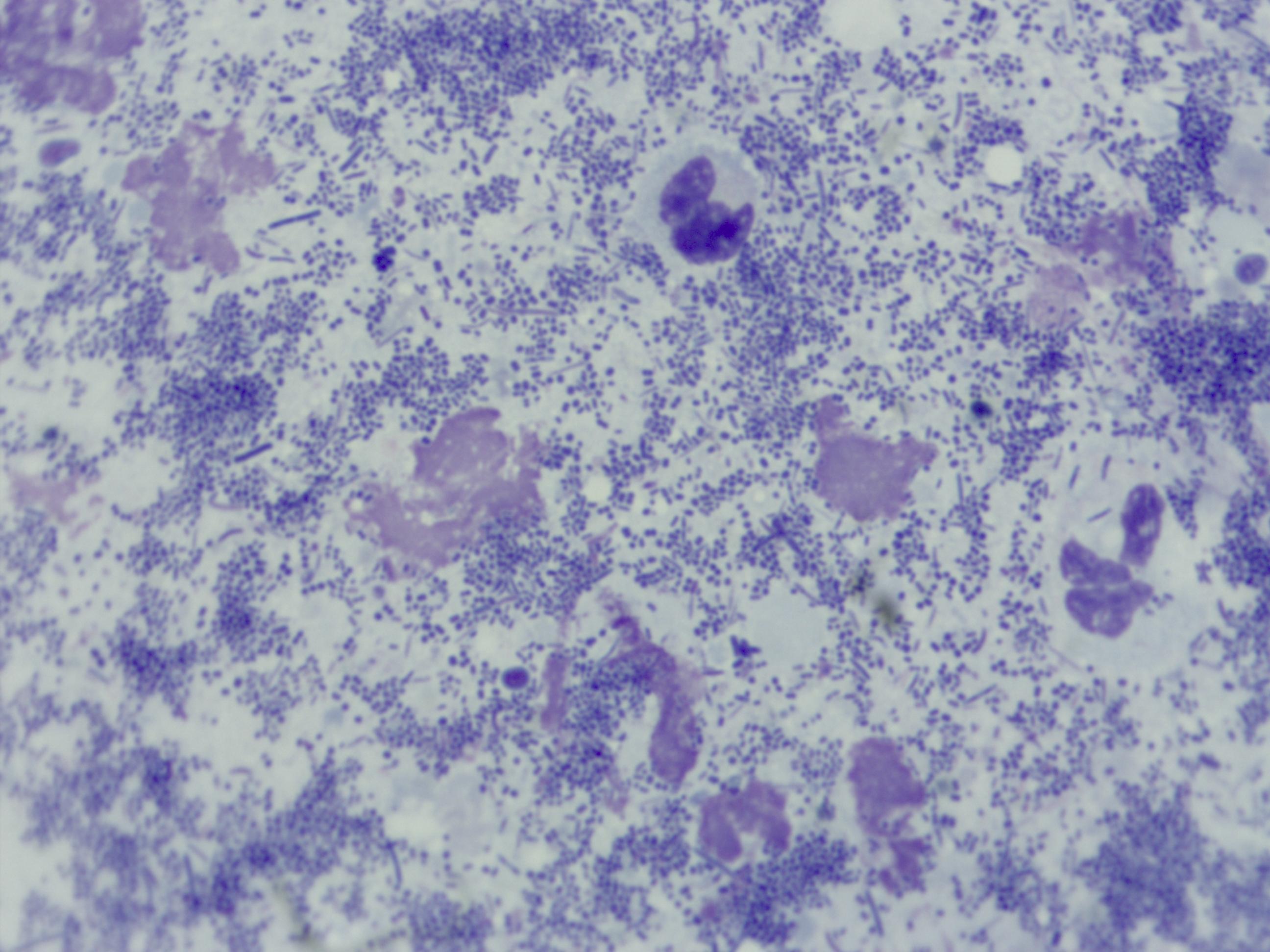
Diagnostic cytology, also called impression smear cytology, is a common diagnostic test that is used to determine the presence of bacteria, yeast or other infections, inflammation and neoplastic (cancer) cells, and alert the dermatologist to other potential disease processes. Samples for diagnostic cytology are often collected by cotton swab, direct smearing of a glass microscope slide, use of clear tape to collect samples, or in some cases fine needle aspirate to obtain samples from a mass or lesion beneath the skin.
By performing this test in-house, veterinary dermatologists can obtain valuable information about a patient or condition while the pet is in the office. This allows for more accurate diagnosis, better decision making on therapies and better patient outcome in managing problem skin diseases.
Skin Scrape
Skin scrapes are another useful tool often performed by veterinary dermatologists. Unlike cytology, skin scrapes are performed by using a dulled scalpel blade to scrape along the surface of the skin to collect material for examination under the microscope. Material is placed onto a microscope slide with a small amount of mineral oil and is examined closely for evidence of parasites. Skin scrapes are used primarily to look for mites including Demodex or the mites that cause Scabies.
Demodex mites commonly live within hair follicles of the skin and may cause inflammation, hair loss scaling and secondary infections. Since these mites live below the surface of the skin, a “deep” skin scrape is needed to identify them. Deep skin scrapes are often performed in relatively small areas (approx. 1 cm diameter) and typically leave a small superficial abrasion, indicating that the scrape has been done deep enough to find the mites.
Superficial skin scrapes involve a broader sweeping scrape done over a larger surface area. This type of scrape is designed to collect surface crust and skin cells and often leave little external evidence that it was performed. Superficial scrapes are used to look for Sarcoptes and other surface mites that may cause inflammation and in many cases intense itching and scratching. Unlike Demodex mites, parasites found on superficial skin scrapes may be elusive with only a few present in a given area making them difficult to find.
Skin scrapes can be a game changer since finding mites can dramatically alter the course of treatment and often leads to much more rapid resolution of skin disease and discomfort.
Trichography
Trichography is the microscopic examination of hairs. Normal hairs of animals have well documented appearance and structure. By plucking hairs from an animal with skin or haircoat disease, a veterinary dermatologist can evaluate the hairs and look for evidence of infection, trauma or malformation. This can also identify parasites like Demodex that live within the hair follicle.
Dermatophyte Testing
Dermatophytosis (ringworm) is a fungal infection of the hair, superficial skin, and occasionally nails. It’s not caused by a worm and often lesions are not in rings. Dermatophytosis is instead caused by infection with one of three species of fungal organism: Microsporum canis, Microsporum gypseum, and Trichophyton mentagrophytes.
Common sources of infection include infected pets, rodents, contaminated environments, and occasionally the soil. While less common than many other skin diseases, dermatophytosis is a contagious and zoonotic disease that can in some cases be transmitted to humans. This makes it an important disease to assess and diagnose when the index of suspicion is high. Methods for diagnosing dermatophytosis include fungal culture, PCR testing and on occasion skin biopsy. In some cases, fungal spores may be identified on impression smear cytology raising the suspicion of dermatophytosis. Woods lamp examination may also increase suspicion of dermatophytosis.
If a veterinary dermatologist feels it is appropriate to test for dermatophytosis, most often he or she will collect hair and skin samples from the area(s) involved for one of these tests. Identification of the specific fungal organism responsible for skin lesions via fungal culture is the most reliable of these methods. Fungal cultures often take 14 to 21 days to complete to allow identifiable fungal organism growth.
When treating a pet for dermatophytosis it is imperative to treat the animal beyond clinical cure, when no further lesions can be seen. To confirm that the infection has been completely resolved a fungal culture is typically repeated to confirm successful therapy.
In-House Blood/Urine Testing
Skin disease in animals can sometimes be an external marker of an internal problem. Examples include disease such as Hypothyroidism, Cushing’s disease and others. For these conditions, information provided by bloodwork or urinalysis can be vital to reaching a diagnosis and directing a proper course of treatment. Animal Dermatology Clinics perform many blood tests in our in-house laboratory, providing more rapid information and diagnosis.
Skin Biopsy and Dermatopathology
To perform a biopsy is to surgically remove a portion of diseased skin, tissue or tumor in order to further analyze this tissue and understand the disease process more completely. For some skin diseases, this is the only accurate way to differentiate one disorder from another and can be the difference between accurate diagnosis and the wrong direction.
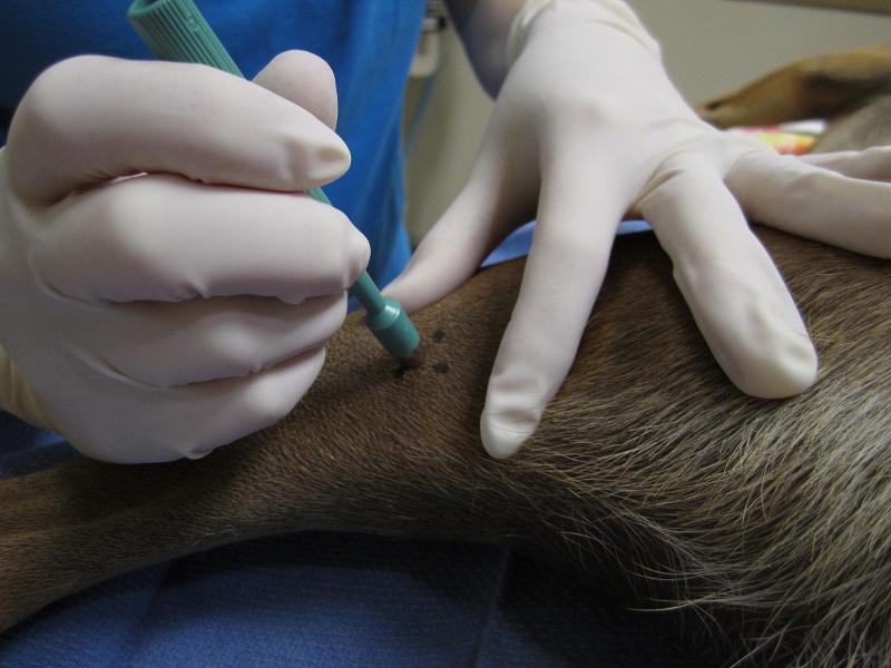
To perform a skin biopsy, most animals are sedated or anesthetized and often a local anesthetic is used to provide further anesthesia and pain control. Many skin biopsies are performed using a “punch” biopsy instrument - a small circular blade that may vary from 4 to 8 mm. Use of a biopsy punch allows a small sample to be obtained from areas of concern and are often sutured with a single stitch.
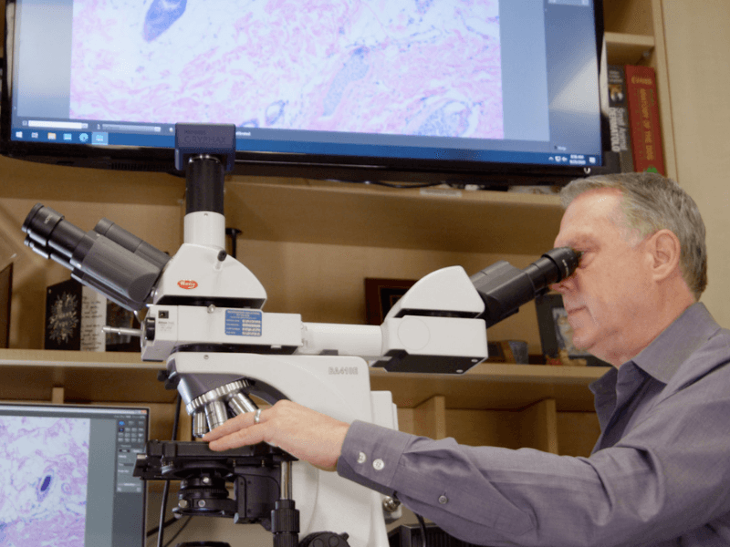
What happens to biopsy skin or tissue after this procedure is performed is of equal importance as the procedure itself. Animal Dermatology Clinic is fortunate to have an in-house dermatopathology service to interpret findings from the biopsy once the tissue has been processed. Dr. Wayne Rosenkrantz, DVM, DACVD, and Dr. Kelly Keating, DVM, DACVP provide dermatopathology interpretation services to both Animal Dermatology Clinic dermatologists, and other specialists and general practitioners. For veterinarians interested in learning more about this service, click here.
Diagnostics - Biopsy 1 - Biopsy procedure Image 2 300dpi[1]-1
In-House Diagnostics-1-1
Diagnostics - Cytology 2 - Bacteria TNTC
Signs and conditions this addresses
Conditions we treat
Common questions
Ready to help your pet?
Request an appointment and our team will guide you through the next steps.